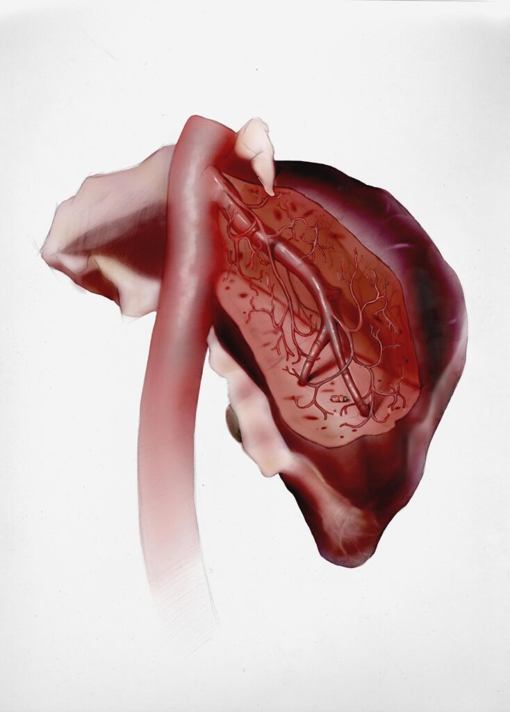
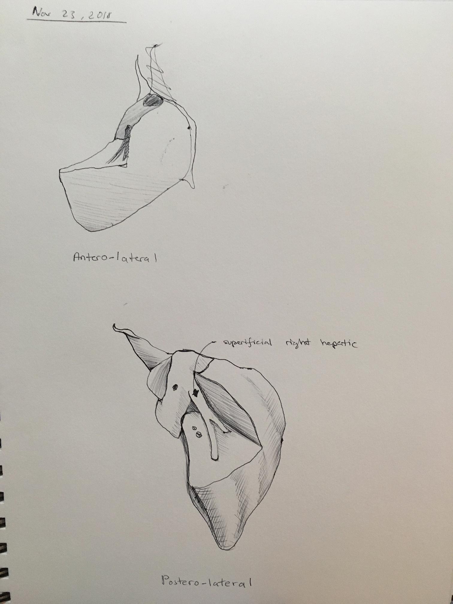
Hepatic Venous Tree of Segment VII (Postero-lateral) Illustration
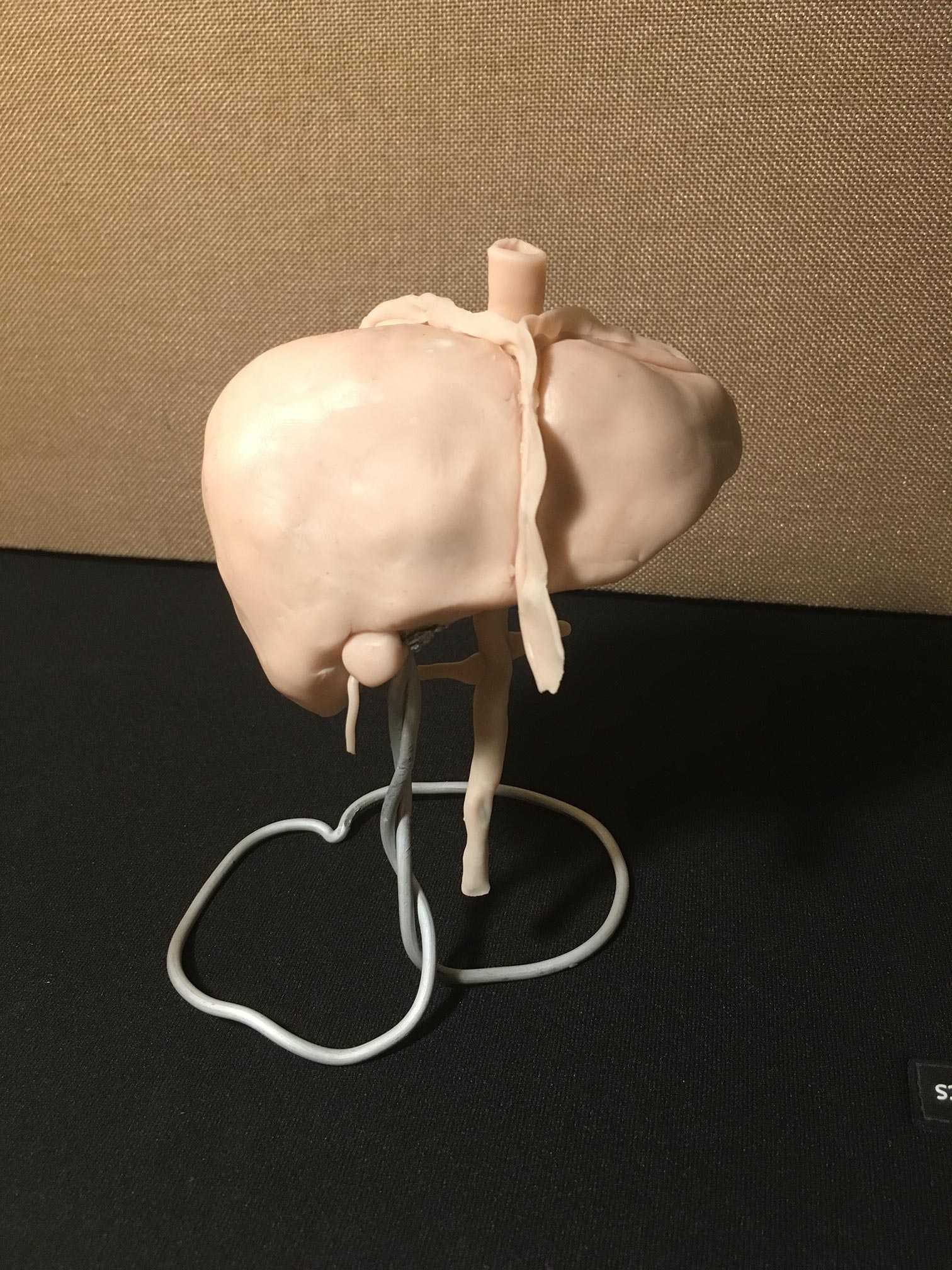
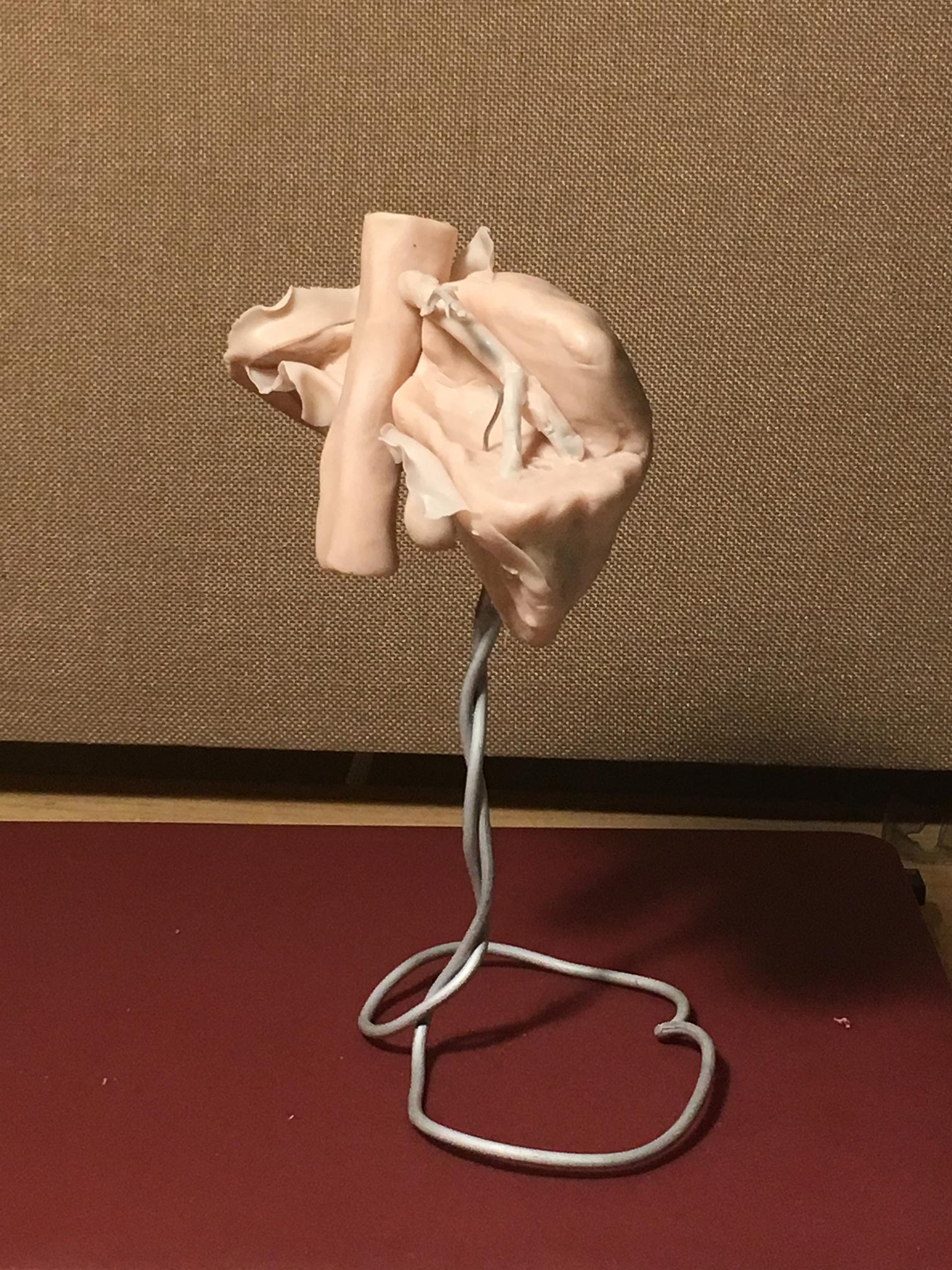
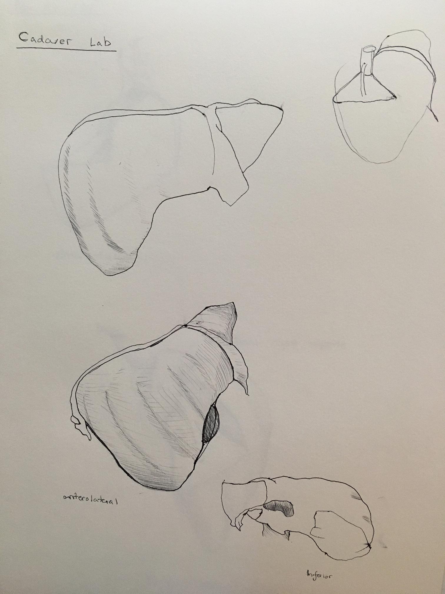
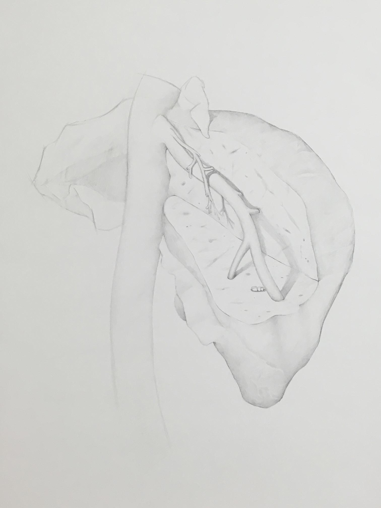
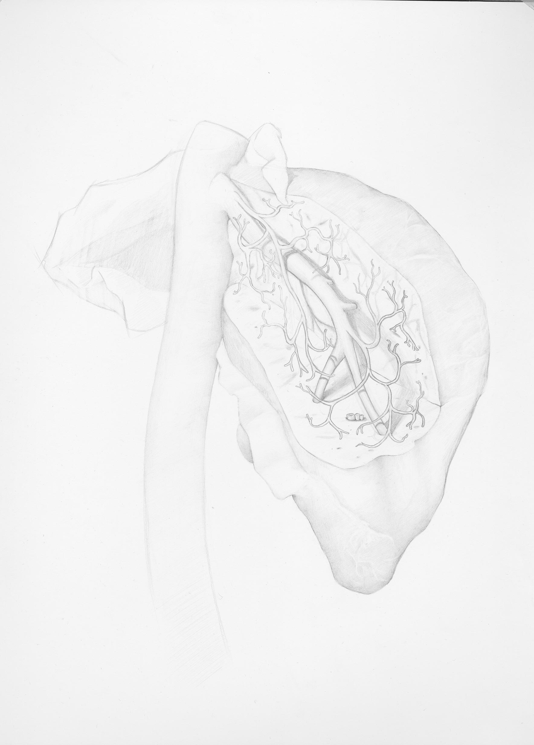
This illustration was created for a biomedical communications course called: MSC2001- Visual Representation of Medical Knowledge. It was an exercise in how to visualize an anatomical area that is challenging to see. In this case, the segments of the liver are divided by which blood supply feeds into them. Thus, the most accurate method to visualize each segment’s boundaries is to ligate arteries in surgery and observe a change in the tissue colour. Since that option was not possible, I relied on research, maquette-building, plastinated venous models, cadaver dissections, feedback from anatomists, and digital 3D models. The final illustration was first done with grisaille technique (i.e., light tonal rendering) with colour applied in Adobe Photoshop afterwards. It was a very useful good exercise in exploring different ways to visualize the “unseeable”.
Objective: To visualize a non-standard anatomical view of the venous system of segment VII of the human liver.